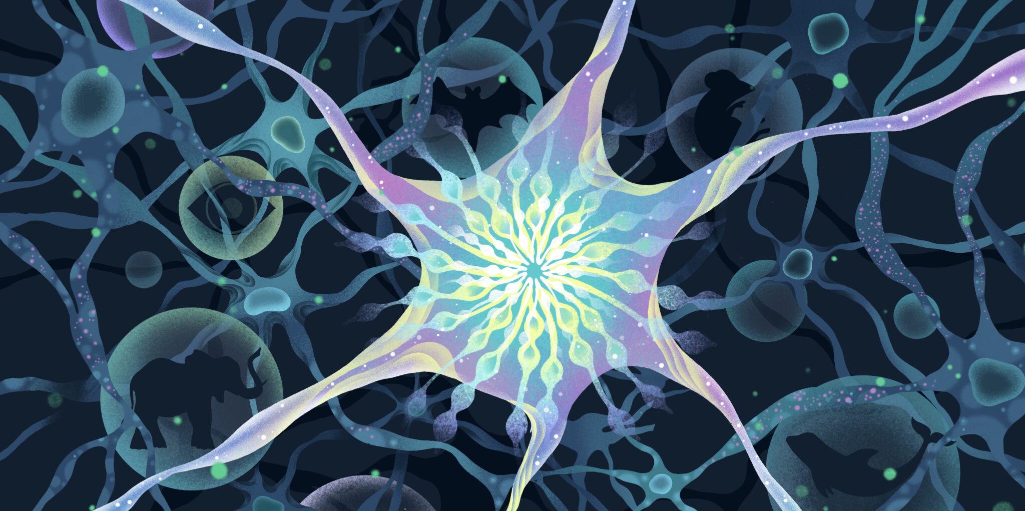
At first glance, Micha Drukker’s lab houses what is possibly the world’s most disappointing zoo. There are naked mole rats, mice, bats, elephants, pilot whales, even a Northern white rhino. But the mole rats are not tunneling, bats aren’t flapping overhead, and there are no large aquariums anywhere. There’s not a rhino horn to be seen.
Drukker’s zoo, at the University of Leiden in The Netherlands, is more wondrous than it first appears. To understand its appeal, one needs to look in the freezer. It is a zoo of cells. Stem cells, to be precise. These cells—induced pluripotent stem cells, or iPSCs—from many different mammal species have vast potential. They could become any cell from their species. The iPSCs could form cells in the gut, which rise up and die within a few days. They could become fat cells, lasting around 8 years. Or they could become neurons, which in the central nervous system can live as long as the organism itself.
Neurons don’t seem to follow the same rules as other cells in the body. They don’t age in the same way, and in human adults, they don’t get replaced. “We know that the majority of neurons, they are not replaced during our lifetime,” says Drukker. “And they have very little regeneration or redundancy.” Poke the tip of a baby’s finger with a pencil, and the signal will travel up sensory neurons to the spinal cord and brain—and potentially make the baby cry. Poke that same finger once that baby is an adult, and the signal will travel the same route, along the very same neurons. The only difference is that an adult will wince instead of wail.
These long-lived cells in our bodies and brains are key to the consistency of our memories and functions. But their own longevity varies drastically depending on species. Mouse neurons live only about two years—as long as a mouse does. An elephant’s neurons, on the other hand, could see seven decades, just like a human’s.
How do these cells live such long lives without ever being replaced? What protects them from life’s hard knocks and allows them to function without breaking down? One clue might be in nuclear structures called paraspeckles. These tiny granules of RNA and protein increase when a cell is stressed. The brains of people with Parkinson’s disease have more of these tiny structures—and even more appear when scientists give neuroprotective drugs. Species such as naked mole rats—a model for an extra-long, if slightly disgusting, life—have different expressions of paraspeckles in their guts than shorter-lived species.
These paraspeckles are one of the reasons for Micha Drukker’s tiny zoo. The mammal species represented have lifespans from only a few years to many decades. By stimulating the iPSCs from these species to form neurons and examining how, when and why their paraspeckles form, Drukker and his colleagues hope to understand how the structures contribute to a cell’s—and an organism’s—lifespan. The results could one day contribute to new treatments and understanding of neurodegenerative diseases such as Parkinson’s, dementia and amyotrophic lateral sclerosis (ALS).
Pinpointing paraspeckles
The scientist who discovered the paraspeckle, the molecular cell biologist Archa Fox, still teaches undergraduate cell biology at the University of Western Australia in Perth. She loves to tell the story of finding the paraspeckle, not to boast, but to show her students how much in the body remains to be discovered. When she was an undergrad, Fox says, “I thought that these were the parts of the cell, and I could draw a picture of an animal cell and what was in it.” She is eager to inspire her students to look deeper. “You don’t know what you don’t know.”
Fox began to learn what she did not know in graduate school in the late 1990s, when she was studying the different proteins inside the nucleolus—a structure inside the nucleus of a cell. The nucleolus is where ribosomes are formed—the cellular factories that take RNA in and pump proteins out. The nucleolus itself is packed with proteins, and Fox’s job was to figure out where they went. She gathered unidentified proteins together, fused them with green fluorescent proteins to make them glow, and followed their paths.
One protein did not follow the rules. “One of these proteins didn’t go to the nucleolus at all, it went to these little dots in the nucleus,” Fox recalls. Her microscope showed tiny glowing dots and long glowing sausages. “I was literally at the point of going, Okay, I’ll put this in the rubbish bin…this protein does not fit, you know, our dogma.”
But Fox did not toss these strange structures in the trash. Instead, she continued to follow the glowing proteins. “It’s an example of serendipity and completely following your nose in science as opposed to having a hypothesis and proving or disproving it,” she says. Tracking the proteins over time showed they only passed through the nucleolus and ended up elsewhere in the nucleus instead.
The proteins weren’t just dots, and they weren’t just proteins. Instead, they were proteins clamping on to a single long non-coding RNA called NEAT1. Each long RNA attracts sets of proteins to it, and forms into a V-shape. The V-shapes then group together with others of their kind at the closed ends of the V, forming tiny microenvironments. As more and more group together, they reorganize, producing long, sausage-like structures, the tiny tubes that Fox saw in her microscope. Fox called these dots paraspeckles—simply because they were near other sub-nuclear structures called speckles—and she showed this new cell feature to the world in 2002.
These were not organelles, the tiny “organs” that most high school students learn about that make a cell function. Organelles are bound by membranes, solid chains of fatty acids. Paraspeckles are not so well defined. Instead, they assemble together without the wall of a membrane—giving them extreme flexibility to form and fall apart as needed.
As they form, paraspeckles create a phase separation—an area that has different properties than the rest of the nucleus, The phase separation can create an area that is more liquid, or it can become more gel-like.
And this might be key to what paraspeckles do in the nucleus. They could be like a holding tank. A sponge. A time-out corner for messenger RNA and the proteins that help produce it. “You were produced, but we don’t really need you to work, so let’s put you somewhere safe,” explains Hermona Soreq, a molecular neuroscientist at the Hebrew University of Jerusalem. Paraspeckles could in this way serve as a control point between the transcription of DNA to RNA and the translation of that RNA into proteins. “If [the cells] put them into the paraspeckles, they can’t…go to the cytoplasm to interact with ribosomes get translated,” Soreq says.
But in this metaphor, the holding tank or sponge is also made of the very molecules in the tank. NEAT1 acts as a seed—like the tiny particles of dust that help raindrops and snowflakes to form. “The proteins then bind onto it, and then it’s like a chain reaction, and they interact with each other,” Fox notes. “It has this kind of transience, if you like, it’s not as fixed as something like a mitochondria.” Some proteins that bind are only parts of a paraspeckle. But other molecules get caught up, attracted by the growing conglomerate.
Less NEAT1, and fewer paraspeckles could mean less attraction, and more RNA and proteins free to do cellular business. But more NEAT1 could mean more tiny little drops of temporarily restrained molecules. In this way, Drukker says, paraspeckles are “a knob in a way that controls the level of protein translation in a cell.” The knob, so far, has been found only in mammals, though related mechanisms may also exist in reptiles. What, then makes those paraspeckles form, or not? What makes the knob turn?
Shorter lives through stress
One of the signals that makes the knob turn could be cellular stress, or cellular change. iPSCs, the absolute base model of cells, do not have paraspeckles. They form when stem cells begin to differentiate into a specific cell types. But then, in neurons, they disappear. “If they are differentiated into neuronal lineage, they downregulate them, and postmitotic neurons don’t have them,” says Tatyana Shelkovnikova, a cell biologist and neuroscientist at the University of Sheffield in England. “So there is a clear sort of stage when they need them for certain reasons.”
All cells with a nucleus can form paraspeckles. Once the cells reach their final maturation destination, whether they do or don’t form the small conglomerates appears to depend on the stresses they are required to weather. Drukker and others have shown that when a cell needs to reprogram—due to differentiating, responding to infection, heat stress, or something else—nucleus size increases, and so do the number of paraspeckles.
Healthy adult neurons have very few paraspeckles, perhaps because there is relatively little to trouble them. They are protected from many infections, and do not need to divide or differentiate once they achieve their final state. When they do form paraspeckles, it is often because something has gone wrong. “We know that a collection of long non-coding RNAs increases in response to stress or brain disease,” Soreq says. The post-mortem brains of people with Parkinson’s, for example, show increased NEAT1 and paraspeckles. “So my guess, it’s right now only a guess, is that the function is needed in conditions of disease, probably to protect the brain from deteriorating even faster than what it does otherwise.” Paraspeckle increases have also been seen in other neurodegenerative diseases such as ALS and Alzheimer’s disease. Outside of the brain, paraspeckles increase in tumor cells exposed to radiation, and may help some types of cancer cell evade treatment. But in other contexts, paraspeckles can help suppress tumor formation.
Are those paraspeckles a sign of cell protection? A knight riding in to save the day? Or are these cells showing high levels of paraspeckles because they are the last gasp of a dying cell? “I think the hardest element here is the chicken and egg aspect to it,” Shelkovnikoa notes. Does the postmortem tissue of people with neurodegenerative diseases have lots of paraspeckles because the cells used them to live? Or are the paraspeckles a sign the cell was doomed?
Some of the answers might lie in Drukker’s tiny zoo. Results from his lab suggest paraspeckles might serve as protectors. So species that live longer—and encounter more stress—might have more of them in their neurons. “One of the good things about paraspeckles and the connection to longevity and lifespan and stress, you can think about the stress as actually a main proxy of longevity,” Drukker says. “The more stress you have, the more problems you have. You age faster.”
This is what Drukker hopes to do—stress out the cells in his collection. With a grant from the John Templeton Foundation, his lab is developing lines of iPSCs from short-lived and long-lived mammals and coaxing each species to form neurons. The lab hopes to characterize the NEAT1 of each species, constructing a paraspeckle family tree.
Then the scientists will lay on the stress. Drukker hopes to compare the stress responses of short- and long-lived species. If long-lived species pump up the paraspeckles, it could suggest they serve as protection. The lab will also use CRISPR gene editing techniques to increase NEAT1—increasing paraspeckle formation without stress, to find out what paraspeckles can do on their own.
The results could indicate what turns the knob on paraspeckle formation, and what turns of the knob signify. If paraspeckle formation is protective—and neurons with more of them live longer—then future drug exploration for neurodegenerative disease could focus on increasing paraspeckles in cells at risk to help them resist stress. If, on the other hand, paraspeckles are a sign of a cell about to give up the ghost, treatments that reduce them could help cells cling on longer.
“This is an exploratory project,” Drukker says. “It’s not just to solve small problems.” Paraspeckles and other membrane-less organelles reveal a method of cellular regulation that scientists are only beginning to understand. With the cells in his zoo, Drukker hopes to explore how these structures work, and what they mean for our bodies as a whole. Exploration into those basic questions, he says, is what really matters. “That’s how you discover really new, fundamental things.”
Bethany Brookshire is an award-winning science journalist and author of the book, Pests: How Humans Create Animal Villains. Her work has appeared in Scientific American, The New York Times, The Washington Post, The Atlantic, and other outlets.