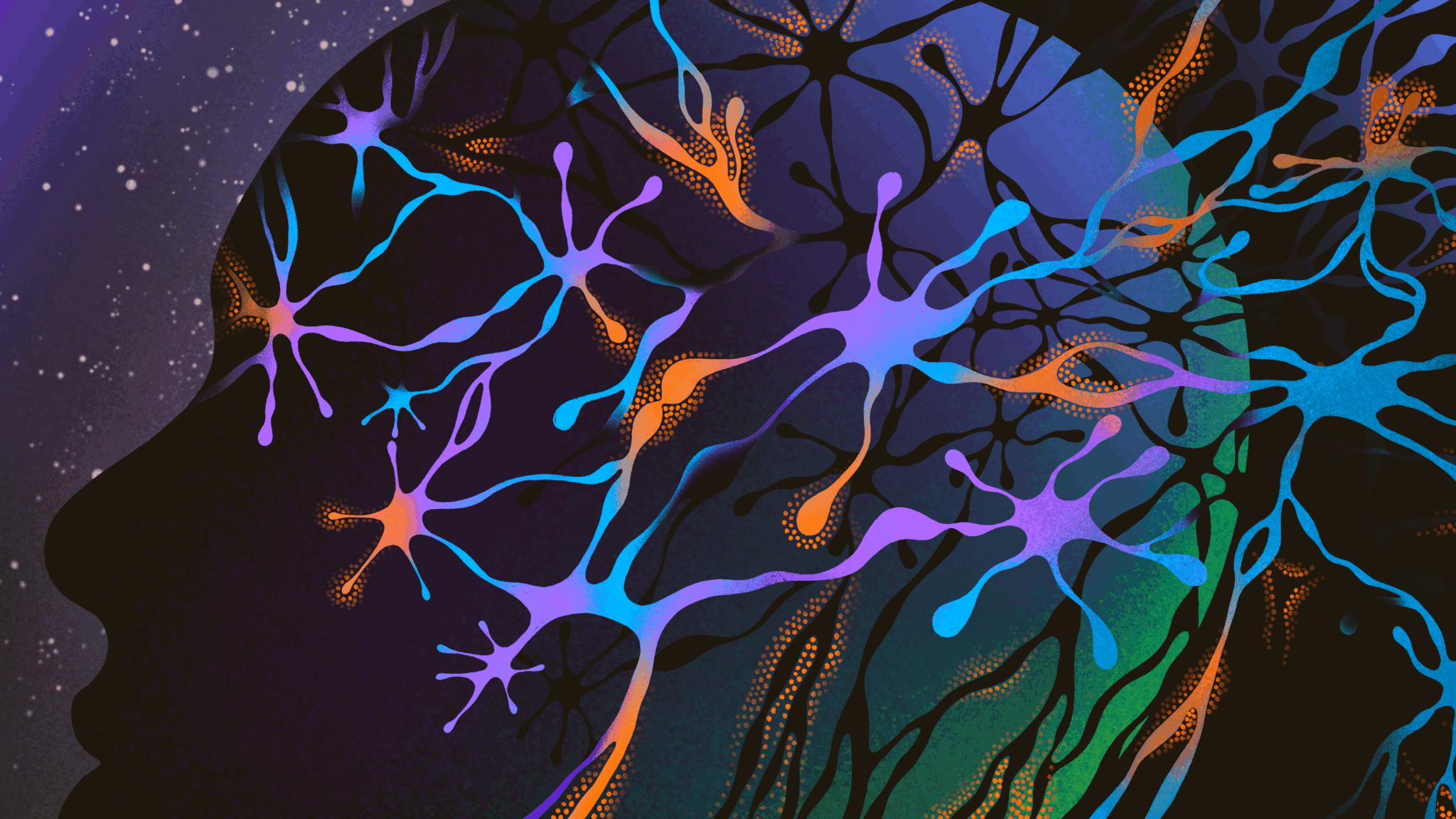
Our bodies are constant hives of communication. Hormones surge through our blood. Ions ebb and flow across membranes, generating electric potentials from one cell to another at blinding speed. Chemicals released from one cell cross tiny gaps to another, passing messages. But when we attempt to imagine this bodily communication, we have to bring our own light. After all, light mostly ends at our skin. It’s easy to assume that the actions inside our bodies are taking place in warm, wet, squishy darkness.
What if we were wrong?
For decades now, scientists have caught flashes of light inside cells. Lentil seeds emit tiny, faint sparks. The mitochondria of cancer cells appear to respond to light. Receptors for light—proteins called opsins—are buried deep in the brain in the hypothalamus, an area that should never, in theory, see the light of day.
All living things, it turns out, produce low, constant levels of photons, simply as a result of cellular activity.
Could cells use this self-created glow? After all, nature rarely produces something without making use of it. For instance, light could function as another kind of messenger in the brain, amping up the speed of communication.
Detecting light produced by neurons is a tricky proposition, and finding out if neurons can use those photons is harder still. With funding from the John Templeton Foundation, physicist Pablo Postigo and neuroscientist Michael Telias at the University of Rochester in New York hope to combine their talents to capture these neuronal fireflies. It’s a risky proposition. But if scientists can show that light in brain cells moves and has a purpose, a whole new world opens. These tiny sparks could contain keys to questions about how neurons control their own firing rates, and perhaps even shine some light on the roots of consciousness.
Get your glow on
Some organisms—fireflies and some comb jellies, for example—can glow intensely, the result of chemical reactions that produce intense luminescence. “Fireflies, those are striking examples of bioluminescence, they emit a lot of light,” Postigo explains. It turns out, he says, “everybody does it, but we cannot see because it’s so weak.”
In this case, “everybody” means everybody with cells—cells performing chemical reactions to produce energy. “Most of the production of photons in a normal natural setup happens in the mitochondria,” explains Telias. As mitochondria run their metabolic reactions to make ATP–the energy currency in many cells–they will also produce reactive oxygen species, or ROS.
Nature lets very little go to waste, and so these small chemicals are far more complex than simple cellular trash.
“They are important signaling molecules,” says Christoph Simon, a theoretical quantum physicist at the University of Calgary in Canada. In planarians (flatworms), for example, these tiny chemicals control growth, he explains. “But they are also aggressive little things.” The tiny molecules of ROS will bind to almost anything, and are implicated in diseases such as cancer due to their ability to bind to and damage DNA.
So we are used to thinking of ROS as negative—people wouldn’t be touting the antioxidant effects of acai berries if they weren’t. But this binding ability could also be the key to lighting the inside of a cell. An ROS can attach to lipids—fatty molecules that make up your cell membranes. The reaction forms a tiny duet of oxygen and hydrogen—a hydroxyl radical. This tiny, hyper-reactive molecule attacks another lipid, forms another radical, and so on. “It’s a thing that keeps going until two of those radical molecules meet,” Simon says. When they do, they react and release radiation.
This radiation is not in the form of heat, though we do produce heat. “Any body of a certain temperature emits radiation as a function of that temperature,” Simon notes. Instead, the energy from the cellular processes can be released as a photon. This is biophoton emission, otherwise called ultra-weak photon emission, or ultra-weak chemiluminescence. “A biophoton per se is just a photon, which is a quantum of light,” Simon says. “It’s called a biophoton when it is emitted by living cells or living systems.”
These photons travel in wavelengths on the nano-scale, with around 470 nanometer wavelengths appearing to our eyes as blue, and 570 as yellow. Each photon is 1000 times smaller than a micron (a single hair is around 100 microns). This makes photons far, far smaller than the comparatively gigantic molecules that fill our cells. Tiny photons traveling in huge megacities of proteins means many biophotons will get lost almost immediately after they are created. “There is a big chance that the vast majority of the photons just scatter,” Telias says. “The question is if a small number of them, a portion of them, can be channeled into a direction, and if that happens, whether that has some kind of physiological consequence.”
The brain’s ‘optic fibers’
If cells can produce light, they might be able to use it.
“I don't think nature does anything without having some reason,” Postigo says.
Photons could just be byproducts, brilliant sparks of metabolic waste. But Telias and Postigo don’t think so.
After all, cells in the eye contain receptors for photons. The retina has a very specific molecular cascade that takes photons and converts them to electrical signals to pass on to the brain. “Cells in the retina can react to light because they are flooded with retinaldehyde,” or retinal, Telias notes. These molecules are linked to receptors called opsins. When photons hit the retinaldehyde, it changes shape, triggering the opsin. “You need to have retinal, you need to have the opsin, and they both need to interact, and the photons need to hit it,” he explains. “All of that machinery is exquisite, and it’s beautiful and it exists in the retina. We don’t have any evidence that that machinery specifically exists elsewhere in the brain.”
In addition, the retina is constantly flooded with light. “The amount of light that hits the retina is immense,” Telias says. “And what we are talking about regarding the production of photons inside a cell somewhere in the brain is the exact opposite. We’re talking about very, very low, very weak emissions.”
Could a cell take these very weak emissions and detect them? Could cells do more, conducting biophotons in streams to serve as messengers? There are indications that it might be possible, particularly in the brain. Brain cells appear to generate more photons than other cell types—photons that are generated as these cells conduct their electrical signals. Neurons also have a structure that could serve as a channel for fluids, ions,and even photons.
Each neuron has a cell body, followed by a long, threadlike axon which ends in an axon terminal. The axon is most well-known for its ability to transmit electrical signals. Ions flow in and out of the cell membrane, sending a wave of signal down the axon, to release chemicals at the end which stimulate the next neuron down the line.
The speed of this electrical activity is immense, but signals move even faster if the axons are myelinated—surrounded by support cells called astrocytes which wrap the axons in fatty myelin sheaths. These allow the electrical signals to bounce quickly along the axons, concentrating at gaps between the myelin linings called nodes of Ranvier.
Those nodes aren’t just electrical concentrations. Other scientists have shown that nodes of Ranvier also produce photons while they are conveying their electrical signals.
Producing light is one thing, conducting it is another, and the structure of the axon might hold the key.
“Axons have the right properties to guide photons,” Simon says. His lab has been building theoretical models of how axons could serve as waveguides. “There’s two things you need, low absorption in your relevant range, but then you need a higher refractive index,” he explains. “That’s how you confine light.” The membranes of axons have both of those things, low absorption, and a high refractive index. “The axon itself has a higher refractive index than the surrounding liquid, but the myelin is even better.”
The two combined could allow streams of photons to move along inside the axon without escaping or being absorbed. Simon’s lab has constructed models accounting for the vagaries of neuron shapes, axon sizes, and the tiny amounts of photons produced in his lab’s models. “Could it still work?” he asked. “Our answer was, yeah, it seems like it could.”
The proof is in the photons
Postigo has always studied the world at the nano-scale, and the structure of axons intrigued him. He noticed that they seemed a bit like optical fibers in their structure and capabilities, and wondered if axons might be able to act naturally in the same ways that manmade optical fibers do. It’s a relatively simple idea—that neurons could produce photons which they could then use as signals—but almost every aspect of it still needs to be proven. Scientists need to catch the waves of light traveling down a cell. They need to find what those photons might hit at the end of their journey. And they need to understand what signals those tiny waves of light might be sending at all.
Postigo and Telias think they know where to start. “Let’s try to see first if we can even detect photons rising in one side and moving all the way to another side,” Telias explains. That part is going to be hard enough.
Postigo’s lab is working on extremely tiny nanophotonic probes. “We use very thin optical fiber,” he explains. These tiny probes will then be tested against fabricated axons before going up against the real thing.
Of course, the probes will need photons to detect. Even in neurons, relatively few of them are produced at any one time, only about 1000 photons per centimeter squared per second—not something the human eye could easily detect. So while Postigo develops probes, Telias is growing neurons that express fluorescent proteins. “We are forcing them to produce a large amount of photons in the nucleus,” he explains.
The goal is to use the probes to detect these artificially produced photons. The trick will be getting the probes to cozy up perfectly to the axon of a neuron. It “should be very close to the axon, but without touching it, because we could damage it,” Postigo explains.
The goal is that if the probes can get close enough, photons will “hop” from the axon to the probe, allowing the researchers to watch in real time as light ripples along a neuron, if it happens at all. “Even if it is a negative answer, it could be useful to know that ‘well, this is not happening,’” Postigo says. “But if a positive answer happens, it will open many, many, other doors.”
An entirely new method of signaling in the brain raises the question—what are those signals doing? Because biophotons are generated by metabolic processes, perhaps they help to regulate those processes, Telias notes. Or perhaps because neurons need to control their firing rates, light could give them quick feedback and help them adjust their electrical potentials. New methods of signaling could also offer new potential answers to big questions like consciousness. “We really don’t know exactly what we’re getting into, but this is just a question of whether this is happening first and foremost,” Telias says.
The research is just beginning, and with so many unanswered questions, the research is high risk. “This is the highest level of risk that a scientist can take, because the answer can be no. There is no evidence that this is happening, and that is a real possibility, right?” Telias says. But “we all want to get the yes.”
Bethany Brookshire is an award-winning science journalist and author of the book, Pests: How Humans Create Animal Villains. Her work has appeared in Scientific American, The New York Times, The Washington Post, The Atlantic, and other outlets.
